Case 19 - A Shocking Twist
Author: Dr Nick Mani Reviewer: Dr Nish Cherian
A 60-year old male presents with palpitations and lightheadedness. ECG shows a narrow complex tachycardia associated with haemodynamic instability.
DC cardioversion is about to be performed but, upon urgent Consultant review, other causes of shock are considered.
You elect to perform a shock POCUS protocol to enhance your clinical assessment: -

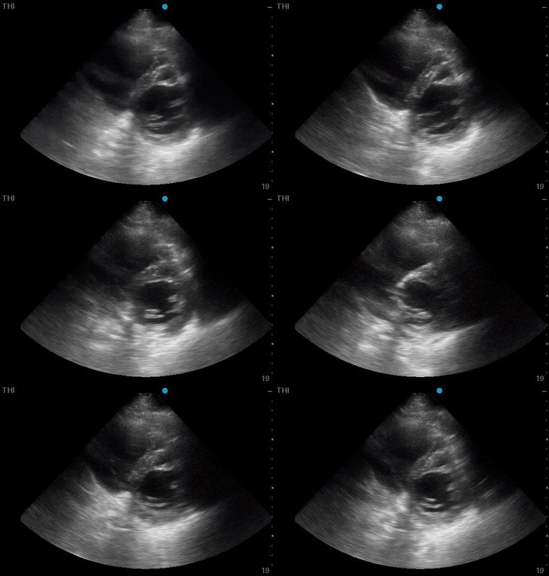


Clips Collection- Relevant clips of the Focused POCUS enhanced clinical assessment*
Panel A- Cardiac (PLAX/PSAX/A4C/SX/SC/IVC & modified PSAX **)
Panel B- Abdominal Aorta (short & long views)
Panel C- Renal (long views)/Bladder (short views)
* Lungs, FAFF, Hepato-Biliary, Appendix, and Proximal Leg Deep Veins were all performed with low suspicion of pathology (Images not shown) as an extended shock protocol
** PLAX- Parasternal Long Axis, PSAX- Parastenral Short Axis, A4C- Apical 4 Chamber, SX/SC- Sub-Xiphoid/Costal
-
Moderate-severe LV systolic impairment.
This could possibly contribute to shock, or could be the primary cause of it depending on the other findings on full assessment
-
AAA measuring 4.67cm
Unlikely as <5cm (rupture risk 0.5-5% per year)
-
Left renal moderate hydronephrosis with complete obstruction of the ureter with ?stones
Most likely this is the primary cause of shock.
-
Septic shock from unilateral complete infective obstructive nephropathy on the background of moderate-severe LV systolic impairment


Images Collection- CT Abdomen/Pelvis with contrast demonstrating left obstructing ureteric calculus and multiple bladder stones, and AAA measuring 4.7cm
Case Resolution
The patient was admitted to the ICU, had an urgent nephrostomy which drained large amount of pus from the left kidney. POCUS-enhanced clinical assessment significantly improved the quality and efficiency of patient care.
Take home message
Cause of shock could be quickly evaluated by a POCUS protocol as a extension of clinical assessment.
Appendix

The Shock Protocol
The black squares are the probe placement with the dotted lines with arrow(s) as sweeping direction. The smaller sized black square- phased array, medium- linear, large- curvilinear